Understanding Blood Smears

Annabel Travers
BVMedSci (Hons), BVM BVS (Hons), DipACVP (Clin Path), MRCVS
Board Certified Clinical Pathologist
The Power of the Blood Smear: A Simple Tool That Tells Big Stories
If there’s one thing I’ve learned during my residency in Veterinary Clinical Pathology, it’s this: never underestimate the humble blood smear. While automated analysers can crunch numbers and spit out results quickly, they can’t capture the whole story. That’s where a smear comes in – it’s a window into the microscopic world of your patient, full of clues and surprises you might otherwise miss.
Why Bother with a Blood Smear?
Analysers are impressive and mighty useful pieces of machinery, but they’re not foolproof. A blood smear gives you the chance to verify results and identify morphological irregularities, which is especially important when things just don’t add up.
- White Blood Cells (WBCs): More Than Just Numbers
- Double-Checking the Analyser: Sometimes, the analysers get confused. Toxic neutrophils might be counted as monocytes, or a reactive monocytosis might be mistaken for a lymphocytosis. Seeing the cells under a microscope clears up the confusion.
- Spotting Left Shifts and Toxic Change: Even in the absence of a neutrophilia, the finding of bands or toxic change signals inflammation, which might otherwise be missed.
- Morphology of Left Shifts: When referring to a ‘left shift’, we mean to say that immature, non-segmented neutrophils have been identified and are circulating. The further left, the less mature the cells are. Myeloid cells will begin as myeloblasts, the earliest form, which then mature through promyelocytes, myelocytes, metamyelocytes, bands, and finally mature neutrophils. This process usually happens in the bone marrow, but when the need for neutrophils increases – for example during active infection – these cells might be released from the bone marrow a little earlier than planned. Depending on the magnitude of inflammation, a left shift could mean the odd circulating band, or in severe cases, circulating myelocytes or even rare promyelocytes. It is important not to confuse these cells with neoplastic cells.

Maturation sequence from myeloblast to mature neutrophils.
- Toxic change: The term toxicity is a bit of a misnomer. When discussing toxicity, we are referring to increased cytoplasmic basophilia, foamy vacuolation and presence of Dohle bodies (aggregates of retained rough endoplasmic reticulum) that neutrophils quite commonly exhibit under strong inflammatory conditions. Toxic granulation is again slightly different, referring to the retention of pink primary granules seen in promyelocytes (see above image) – a rare sign of very severe inflammation. Don’t confuse these with the pale pink staining ‘secondary granules’ seen in normal mature neutrophils (they are called granulocytes for a reason!)

High numbers of neutrophils in a blood film. Neutrophils have foamy basophilic cytoplasm (toxicity) and reduced nuclear segmentation (bands).
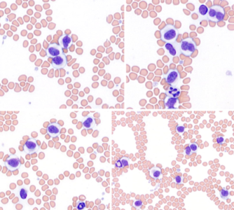
- Catching Atypical Cells: Some of the most fascinating cases come from spotting atypical cells. For instance, with or without a lymphocytosis, an increase in the proportion of small granular lymphocytes could point to developing chronic lymphocytic leukaemia (CLL), which you might not know about without peeking at the film. Likewise, myeloid precursors can only be spotted down the scope, and in the eyes of an analyser, rare lymphoblasts may masquerade as monocytes.

Low numbers of these cells were found to be circulating, even though the total WBC was within reference interval. The nucleus is slightly larger than the neutrophil next to it and contains several pale nucleoli. The patient was known to have lymphoma.

Two atypical large lymphocytes are seen next to a small lymphocyte. A neutrophil and a platelet is also present.
- Platelets: More Than Meets the Analyser
- Clumps Happen: Platelet clumping can lead to falsely low platelet counts, when in reality there are plenty of platelets – this is especially common in cats. Assessing the blood film for platelet clumps is quick and easy, and a manual minimum estimate can be performed at the same time.

Platelet clumps at the feathered edge of the smear.
- Platelet Count: Without clumping, most cats and dogs will have around 10-30 platelets per 100x field of view. To perform a manual count, the average number of platelets in ten 100x fields of view should be counted and an average calculated. This number is then multiplied by 15,000 to get the number of platelets per microlitres of blood.
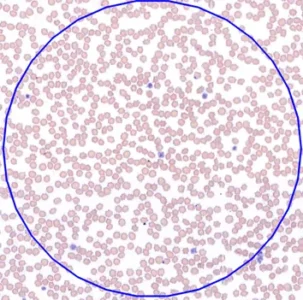
There are 7 platelets in this 100x field of view
- Macroplatelets and Breed Quirks: Some breeds, like Cavalier King Charles Spaniels, naturally have larger platelets due to hereditary traits. A smear can confirm this and prevent unnecessary concern.
- Parasites: A very lucky pathologist might spot a morula from Anaplasma platys lurking in a platelet – a discovery no machine will make for you. This is something to consider in our well-travelled canine patients.

Platelet clumps
- Red Blood Cells (RBCs): The Subtle Storytellers
- Signs of Regeneration: Spotting polychromasia and nucleated RBCs on a smear is a sign that the bone marrow recognises the need for more RBCs and is trying to keep up with demand.
- Hypochromia: The MCHC value is a calculation derived from HCT and Hb, which are prone to error, and is also lower when increased numbers of reticulocytes are present. Confirming hypochromia (and a microcytosis) on a blood smear will help you know when to be worried about genuine iron restricted haematopoiesis.

Increased central pallor suggesting hypochromia. Occasional reticulocytes are also present.
- Shape and Size Matter: A smear can reveal changes such as hereditary macrocytosis in a poodle or microcytosis from iron deficiency. Other changes such as Heinz bodies (denatured precipitated haemoglobin), spherocytes (usually associated with IMHA in dogs) or schistocytes (fragmented due to shear trauma with many possible causes) may also point to pathology requiring exploration.089

An erythrocyte impacts a fibrin strand under flow conditions, forming a schistocyte and a micospherocyte. Image adapted from Harvey’s Veterinary Haematology
- Parasites and Bacteria: A smear may be key to identifying parasites or bacteria hiding out in or on RBC’s. Haemotrophic mycoplasmas and babesiosis are two such examples that can result in severe anaemia in cats and dogs.

Haemoproteus parasite in the erythrocyte of an owl. Parasitised birds rarely show serious disease and are therefore subclinically infected.
Making the Most of a Blood Smear
Smears are simple, but not all are created equal. If you want your smear to tell its full story, here are a few tips:
- Fresh is Best: Time takes its toll on cell morphology. Smears made from aged blood can make cells look toxic or vacuolated, so we prefer to assess morphology from fresh submitted smears. It is also important to ensure smears are not refrigerated or exposed to excessive heat.
- Capture the Feathered Edge: This is necessary for identifying platelet clumps and large neoplastic cells or parasites.
- Perfect Your Technique: A clean, thin smear with an evenly spread monolayer is best. DiffQuik if reviewing in house or send unstained to your laboratory.

The Challenges of Blood Smears (and Why They’re Worth It)
Let me be honest: blood smears aren’t always easy. Even now, I sometimes struggle to identify subtle toxic changes in neutrophils or decide if a lymphoid population is reactive or neoplastic. It’s humbling. But it’s also where the learning happens.
One thing that helps is correlating findings with the patient’s story. For example, a young dog with a lymphocytosis and small cells on the smear probably doesn’t have CLL -context matters. And when in doubt, talking things over with colleagues is a lifesaver and a great learning opportunity. Follow-up tests like flow cytometry can confirm or refute your suspicions, giving you more confidence next time.
A moderate lymphocytosis consisting of small to medium lymphocytes with oval to indented nuclei and expanded amounts of pale blue cytoplasm. This was interpreted as a monocytosis by the in-house analyser. Flow cytometry confirmed T-lymphocyte origin, with moderate loss of the pan-lymphocyte marker CD45 supporting a leukaemic phase of T-zone lymphoma.
Why We Love Blood Smears
There’s something deeply satisfying about finding an answer hidden in a blood smear. It’s like solving a puzzle no one else even knew was there. And while I know I have a long way to go before I feel like an expert, every case teaches me something new.
So, the next time you’re drawing blood, consider adding a smear to the mix. It’s a small step that can make a huge difference.