Smart Diagnostics Part 2: AI to Improve Prognostic Accuracy in Ki67 staining of Canine Cutaneous Mast Cell Tumours

David Walker
Board Certified Anatomic Pathologist
Cutaneous mast cell tumours (MCTs) are amongst the most common malignant skin tumours in dogs. Their biological behaviour can be variable and as a consequence the prognosis can be unpredictable. Whilst well-established grading systems have been developed and are routinely used to assess these tumours, namely the ‘two-tier’ (Kiupel et al 20I0) and older ‘three-tier’ (Patnaik et al 1984) grading systems, these do not correctly predict clinical outcomes in all cases. A subset of MCTs, identified as low/intermediate grade examples (Figure 1) using these grading schemes, do not act as indicated by the histologic grading. This subset of tumours can display more aggressive behaviour, including recurrence, nodal metastasis (Figure 2) and distant metastasis, ultimately resulting in euthanasia or death of some of the affected patients.
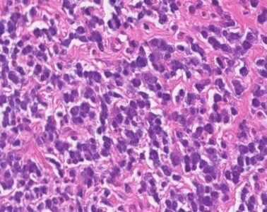
Figure 1. Typical histological appearance of a Grade II/low grade mast cell tumour, exhibiting well-differentiated mast cell amongst a fibrous stroma, with accompanying eosinophilic inflammation.
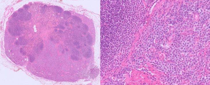
Figure 2. Image of a nodal metastasis of a well-differentiated MCT. Neoplastic mast cells are seen to fill sinuses and efface the nodal architecture.
Multiple additional prognostic markers have been investigated to more precisely predict the prognosis of canine cutaneous MCTs. These tests, such as the Ki67 index, may help to identify those tumours likely to be in the aggressive subset of apparently histologically ‘low/intermediate’ grade MCTs.
What is the Ki67 index?
The Ki67 index is the most commonly requested prognostic marker for mast cell tumours and melanocytic neoplasms. It essentially measures the proportion of tumour cells that are actively dividing in a given snap shot of time. Elevated expression of Ki67 in mast cell tumours has been shown to be statistically associated with more aggressive tumour behaviour and poorer patient outcomes (Scase et al 2006; Maglennon et al 2008). However, formulation of this index typically requires manual counting of large numbers of individual mast cells under the microscope – a method that is inherently time-consuming. Other cells, including stromal cells and inflammatory cells, can also display positivity for this marker, adding to the complexity of interpretation of the test. At VPG, Ki67 stained slides are interpreted by anatomical pathologists experienced with the methodology and analysis. For inexperienced pathologists interpreting Ki67 stained sections, the test can be time consuming, and potentially susceptible to interobserver and intraobserver variability.
To address these limitations, a recent VPG study explored the use of artificial intelligence (AI)-assisted analysis to automate Ki67 cell counting in canine cutaneous MCTs. The goal was to increase efficiency, reduce interobserver and intraobserver variability, and ultimately support more accurate prognostication for clinical practice.
Development of the AI-Assisted Tool
The study utilised QuPath, a platform for digital pathology image analysis (Bankhead et al 2017). VPG pathologists trained a machine learning algorithm to identify and distinguish between Ki67-positive and Ki67-negative mast cells, as well as other neighbouring cells including inflammatory leucocytes, which may complicate the interpretation of the immunohistochemical staining (Figure 3).
Training was conducted using a dataset of 244 representative MCT images, allowing the machine to learn the relevant digital and microscopic features of positive and negative staining (this could include, for example, staining intensity, ‘roundness’ of the cells and other colour/shape parameters). To evaluate its performance, the trained AI system was tested on a separate set of 77 images, which were also reviewed manually by three board-certified veterinary histopathologists. This allowed for direct comparison between ‘machine’ and human in terms of accuracy and reproducibility.
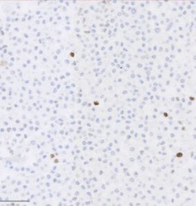
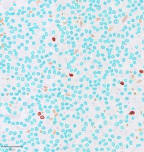
Figure 3. Example of a Ki67-stained mast cell tumour (left). Dark brown round cells are considered to be positive mast cells in this image. A machine-interpreted image (right) highlights positive mast cells (red outline) and negative mast cells (blue outline). Other cells include stromal cells and eosinophils.
Key Findings
The AI-assisted method demonstrates good levels of accuracy, closely matching the results obtained by manual cell counting. Importantly, the automated system significantly reduces the time required to complete the analysis. It also allows for greater consistency across samples, helping to minimise interobserver variability. Remarkably, the time required to complete an image analysis is now less than 1 second, rather than the average time of a ‘manual count’, which is often several minutes.
These findings demonstrate that AI can be a practical advancement in the diagnostic workflow of veterinary pathology providers, particularly for specific tasks that are labour-intensive and potentially subject to interobserver and intraobserver variation. While AI does not aim to replace the expertise of a trained pathologist, it can serve as a valuable aid, particularly in high-throughput and laborious settings, or when standardisation is a priority. Incorporating this AI tool into diagnostic pathology offers benefits for both pathologists and clinicians; More efficient and reproducible Ki67 scoring allows for improved prognostic assessments.
Conclusion
The study aimed to validate the use of AI as a tool to assist with Ki67 cell counting in canine cutaneous mast cell tumours. Whilst traditional manual cell counting to attain a Ki67-index provides clinically useful prognostic information, AI-assisted counts enhance the process with more rapid and reproducible counts, reducing the physical ‘burden’ of a manual cell count. Whilst offering significant improvements in efficiency, all AI-assisted counts are verified carefully by a board-certified pathologist to ensure their accuracy.
References
Bankhead, P., Loughrey, M.B., Fernández, J.A., Dombrowski, Y., McArt, D.G., Dunne, P.D., McQuaid, S., Gray, R.T., Murray, L.J., Coleman, H.G. and James, J.A., 2017. QuPath: Open source software for digital pathology image analysis. Scientific Reports, 7(1), pp.1-7.
Kiupel M, Webster JD, Bailey KL, et al. Proposal of a 2-Tier Histologic Grading System for Canine Cutaneous Mast Cell Tumors to More Accurately Predict Biological Behavior. Veterinary Pathology. 2010;48(1):147-155.
Patnaik AK, Ehler WJ, MacEwen EG. Canine cutaneous mast cell tumor: morphologic grading and survival time in 83 dogs. Vet Pathol. 1984 Sep;21(5):469-74.
Maglennon GA, Murphy S, Adams V, Miller J, Smith K, Blunden A, Scase TJ. Association of Ki67 index with prognosis for intermediate-grade canine cutaneous mast cell tumours. Vet Comp Oncol. 2008 Dec;6(4):268-74. doi: 10.1111/j.1476-5829.2008.00168.x. PMID: 19178685.
Scase TJ, Edwards D, Miller J, Henley W, Smith K, Blunden A, Murphy S. Canine mast cell tumors: correlation of apoptosis and proliferation markers with prognosis. J Vet Intern Med. 2006 Jan-Feb;20(1):151-8. doi: 10.1892/0891-6640(2006)20[151:cmctco]2.0.co;2. PMID: 16496935.